

HIP JOINT REPLACEMENT
It is the biggest ball-and-socket joint. The attachment is framed by the hip bone socket of the pelvis bone. The ball is the femoral head, which is the upper end of the thigh bone.
A smooth tissue called articular Cartilage, lines the surfaces of the joint henceforth empowering them to move easily and effectively. Synovial layer encompasses the hip joint, which secretes synovial liquid that greases up the joint. Tendons and case associates the ball to the attachment and give soundness to the joint.
Hip Joint Fracture And Fixation
PRE OP EVALUATION
- Clinical examination.
- X rays of the knee joint.
- Blood investigations
- CUE.
- Chest x ray.
- ECG.
- 2D echo.
- Physician and cardiologist opinion.
- Anaesthetic check up.
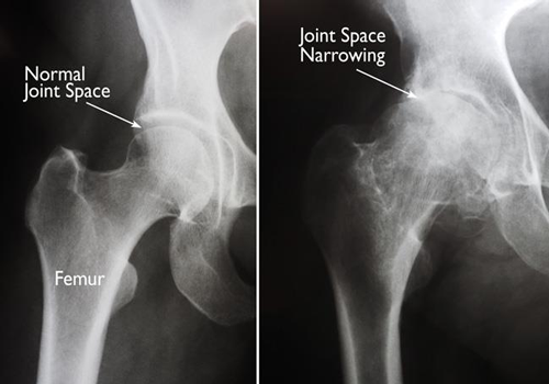
CAUSES OF HIP PAIN
- OSTEOARTHRITIS
- RHEUMATOID ARTHRITIS
- POST TRAUMATIC ARTHRITIS
- AVASCULAR NECROSIS
- CONGENITAL HIP DISLOCATION
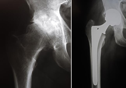
HIP REPLACEMENT PROCEDURE
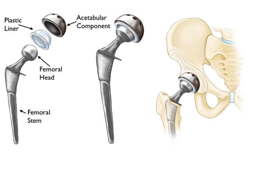
The harmed femoral head is evacuated and supplanted with a metal stem. The femoral stem is either established or uncemented into the bone.
A metal or Ceramic ball is set on the upper piece of the stem.
The harmed Cartilage surface of the attachment (hip bone socket) is evacuated and supplanted with a metal attachment that is verified with screws or bond.
A plastic, Ceramic, or metal spacer is embedded between the new ball and the attachment to take into consideration a smooth skimming surface.
POSTOPERATIVE PERIOD
Patient is made to stroll with assistance of walker by the physiotherapist.
Knee scope of movement and fortifying activities are educated amid the medical clinic remain.
Hospitalization is for 3-4 days.
It will take a month and a half to get back to normal activites and walk without assistance depending on patient capability.